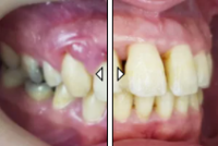
Most Severe Case Of Calculus I Treated. Read The Story Behind The Case.
During my Periodontology specialisation in Bern University, Switzerland I had the opportunity to work on a wide variety of extreme dental cases. This particular case was one of the most severe cases of calculus formation I received. The patient was financially limited and as you can see after treatment the results were great. However, a simple education in brushing from an early age and/or visits to a dentist could have prevented this in the first place! Here is the full story….
In dentistry, calculus (also known as tartar) is a form of hardened dental plaque. It is caused by precipitation of minerals from saliva and gingival crevicular fluid in plaque on the teeth. This process of precipitation kills the bacterial cells within dental plaque, but the rough and hardened surface that is formed provides an ideal surface for further plaque formation.
I previously described how receding gums ruins your gums and smile eventually leading to tooth loss . Calculus build up is just one of the causes of this which can be clearly seen in the pictures below. Actually on removal of the calculus “block” you will note some teeth are missing, gum is destroyed and remaining teeth are left with 5 mm recessions.

Extreme Calculus

Extreme Calculus
The patient, a farmer, 60 years old was living in a remote mountain village in Switzerland. The calculus had built up over time as he never visited a dentist and never brushed his teeth. The calculus block broke and now he was extremely concerned about his health, with little idea as to what was growing in his mouth.
He came for first aid and was treated immediately with extractions, root planning and scaling. The two pictures show the results after the initial first aid treatment. Even though the damage left behind was clear the calculus was gone and the rehabilitation phase could begin. Also the patient was calmed leaving the clinic with a clear understanding of next steps. The patient was rescheduled for further treatment after one week.

Damage Left From Calculus (Lower Jaw)

Damage Left From Calculus (Upper Jaw)
In the removed calculus block (below) you can even see part of tooth #31 embedded in the top left corner. Probably there were parts of the other adjacent teeth also. The size of the block was approximately 15cm x 10cm 10cm. Imagine living with a block that size in your mouth!
The patient received a temporary removable denture as an initial phase. See figure below.
After three months I recommended implant rehabilitation in the frontal lower arch and fixed or removal prostheses, then a visit to the hygienic every three months.

Removed Calculus "Block"

Temporary Denture
The X-Ray below demonstrates the bone loss around the molar teeth and in the lower jaw we can observe radiopacity between 33 to 45 due to the calculus rock located in the floor of the mouth.

Calculus X-Ray
Subsequently, the patient received a “little” lesson in oral hygiene and keeps his new smile well maintained.


