Soft Tissue Augmentation Following Extraction & Socket Preservation
This article is about anterior tooth fracture and the treatment of the soft and hard tissues.
17.93% of extracted teeth are due to root fracture. Most extracted teeth for root fracture were teeth with prosthetic restorations (Popescu SM 2017).
Extracted Tooth with 6mm Aveola Process
Video: Full (Edited) Surgical Protocol Including Prosthetic Preparation, Extraction, GBR & Closing Suturing .
For fractured teeth, the treatment of choice is extraction. However, when we extract an anterior fractured front tooth it usually comes out with the alveolar process, which reassures even more of a bone loss and as a result a compromised bone heal. Due to compromised bone heal a compromised soft tissue healing is expected which could lead to a major esthetic problem.
A healthy beautiful woman with a supragingival smile steps into my office. Her main complaint was mobile crowns in the front teeth (11-21).
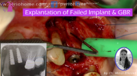
The dental history reveals – this zirconia bridge and posts (11-21) were cemented 5 years ago. The clinical examination reveals a 6 mm mobile fracture at the buccal site attached to the alveolar bone process. The CBCT showed the same (Picture 1).
Another important input to be considered was the patient supragingival smile – making s this case very sensitive. Planning and deep understanding of the details is a must in such a case to ensure the best results.
The treatment was divided into prosthetic and surgical parts.
In the prosthetic part – several impressions were taken
- Pick-up impression of the old crowns
- Impression of tooth 11 with plastic post – indirect post-measurement
- Antagonist and occlusal bite
The surgical part includes:
- Gentle Extraction with Luxators (picture 2)
- taking subepithelial connective tissue graft (SCTG) from the palate and sutured it in the inner side of the gingiva on the buccal side.
- Then a collagen sponge was shaped and inserted at the buccal side.
- Allograft bone was inserted into the socket (picture 3) and then suturing with Nylon suture material (60).
Adding Subepithelial Connective Tissue Graft to the socket preservation is essential in order to bring back the gingiva-lost volume.
SCTG – bed preparation needs to cover the dehiscence part, and to extend laterally from side to side to get the blood supply from the underlying tissues. Ensuring the stability of the graft, by suturing, is a key factor for the success of graft implantation.
Immediately after surgery the bridge 11-21 with reduced pontic (21) contact with the tissue was cemented with temp bond (picture 4). And the patient was released home with adequate instructions.


